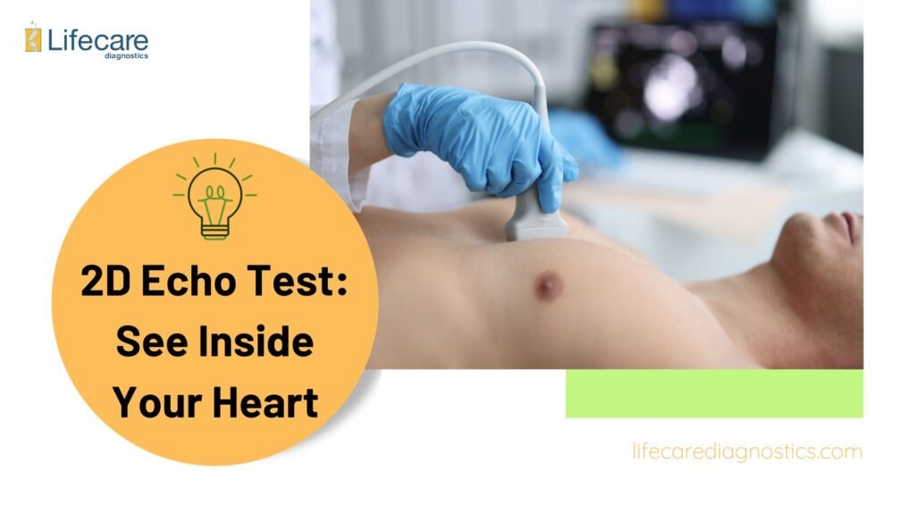
By producing a real-time image of the framework and function of the heart, a 2D Echo test, also related to as echocardiography, offers extensive wisdoms into your heart health. Using this non-invasive test, medical qualified can estimate the size, shape, and function of the heart’s chambers and valves additionally seeking for anomalies like chronic heart defects, valve troublesome, and weakening of the heart’s muscle. It also measures cardiac output, assesses blood flow patterns, and looks for any hints of heart harm or illness.
What is a 2D Echo Test?
A beyond surgery imaging technique used to estimate the structure and function of the heart is known echocardiography, or 2D Echo testing. It utilizes ultrasonography innovation to create real-time images of the heart’s chambers, valves, and blood flow patterns.
The beyond surgery ultrasound of the heart imaging procedure known as a 2D Echo test, or echocardiography, estimates the anatomy and physiology of the heart. It provides images of the heart’s chambers, valves, and blood flow patterns in true time. Diagnoses for heart states such as anomalies in the heart muscle, valves, or chronic faultsare aided by this test. When someone displays heart disease symptoms or as part of a routine cardiac screening, doctors might advise it. In sequence to properly monitor and handle cardiac health, a 2D Echo test is essential for assessing heart function.
Benefits of a 2D Echo Test
An echo test, also called a 2D echocardiogram, is a non-invasive medical procedure that uses sound waves to produce images of the heart. By offering costless insights (cost-effective might be a better term) into the composition and operation of the heart, this diagnostic tool facilitates the identification and treatment of a broad range of cardiac disorders.
Key Benefits of a 2D Echo Test:
One of the main benefits of a 2D echo test is its capacity to estimate the heart’s overall health, involving the function of its chambers, valves, and blood flow. This comprehensive assessment aids in diagnosing various heart conditions, including:
- Heart Valve Disorders: The test can identify abnormalities in the heart valves, such as stenosis (narrowing) or regurgitation (leakage).
- Congenital Cardiac Defects: 2D echo can detect birth defects in the heart structure present since birth.
- Cardiomyopathy: This test helps diagnose weakened heart muscles affecting pumping efficiency.
- Pericardial Diseases: It can detect inflammation or fluid buildup around the heart (pericardium).
Furthermore, the test can assess the heart’s pumping function, also referred to as the ejection fraction, which is crucial in diagnosing conditions like heart failure.
Real-Time Visualization for Enhanced Diagnosis:
Additionally, a 2D echo test provides cardiologists with the capability to view the heart’s movement in real time. This real-time visualization aids them in identifying:
- Tumors or Blood Clots: The test can detect the presence of tumors or blood clots within the heart chambers.
Irregularities in Heart Rhythm: It may identify arrhythmias, which are irregular heart rhythms due to faulty electrical signals.
Conditions Diagnosed with 2D Echo Test
An effective diagnostic tool for identifying diverse heart states is a 2D echo test, also known as echocardiography. Initially, it is capable of detecting congenital cardiac defects, or structural inconsistency like malformed valves or scars in the septum that are present from birth. A 2D Echo test also deputies in the diagnosis of coronary artery disease by estimating coronary artery blood flow and identifying any constriction or blockage.
Additionally, by assessing the sizes, form, and functionality of the heart chambers, this test advice diagnose cardiomyopathy, a class of chaos that impact the heart muscle. Additionally, if pericardial effusion is not treated, it can outcome in cardiac obstacle due to an abnormal build-up of fluid surrounding the heart. This condition is detected by this method.
- Detection of Congenital Cardiac Defects: Identifies structural abnormalities present from birth, such as malformed valves or septal scars.
- Diagnosis of Coronary Artery Disease: Estimates coronary artery blood flow and detects blockages or constrictions indicative of coronary artery disease.
- Assessment of Heart Chambers: Evaluates sizes, shapes, and functionality of heart chambers to diagnose cardiomyopathy, a disorder affecting heart muscle.
- Identification of Pericardial Effusion: Detects abnormal fluid accumulation around the heart, which if untreated, can lead to cardiac complications.
What to Expect During Your 2D Echo Test
You can anticipate a relaxed environment in a clinical setting for your 2D Echo test. Throughout the procedure, a skilled technician will lead you and make sure you’re comfortable and safe. Next, they will take pictures of the composition and operation of your heart using a transducer device. In sequence to get clearer images, you might be asked to shift situations or hold your breath for a time.
Breakdown of a 2D Echo Reports
- Detailed Heart Evaluation: The report provides in-depth details about the structure and function of your heart as seen in the images captured during the test.
Interpreting the Results:
- Normal Findings: An ideal report indicates that your heart chambers, valves, and overall function are within normal ranges. This signifies the absence of any structural abnormalities or signs of dysfunction.
- Abnormal Findings: An abnormal report may suggest potential issues like:
- Cardiomyopathy (weakened heart muscle)
- Coronary artery disease (blockages in heart arteries)
- Congenital heart defects (birth defects in heart structure)
- Abnormal heart valves (stenosis or regurgitation)
- Areas of Concern: The report may highlight specific areas requiring further evaluation, such as:
- Lower ejection fraction (reduced pumping strength)
- Unusual blood flow patterns
Follow-Up and Management:
- Consultation with Cardiologist: Your cardiologist will review the results with you, discuss any concerns, and recommend further steps.
- Treatment Options: Depending on the findings, additional testing or treatments may be suggested to address any identified issues and improve your heart health.
- Importance of Follow-Up: Regular follow-up with your healthcare provider is crucial to ensure a thorough assessment and appropriate management plan based on your 2D echo test results.
Frequently Asked Questions
Is 2D echo better than ECG?
2D Echo provides detailed images of heart structure, while ECG records heart’s electrical activity; both serve different purposes.
Does echo test show blockage?
Echo test can show signs of blockages indirectly through assessing blood flow and heart function.
Is my heart OK if echo is normal?
A normal Echo test doesn’t guarantee overall heart health, but it’s a positive indicator.
Which test is best for heart?
The best heart test depends on individual symptoms and medical history; consult a cardiologist.
Which test confirms heart blockage?
Angiography confirms heart blockage by visualizing blood vessels directly.
Which test is better TMT or 2D echo?
TMT evaluates heart’s response to exercise, while 2D Echo provides structural details; both have their merits.
How much does a 2D Echo Test cost?
2D Echo test prices in Mumbai vary; consult diagnostic centers for accurate pricing.
Recent Blogs:

The Cost of Prenatal Testing: Sympathetic Your Options

Navigating Pregnancy: Positive Double Marker Test Explained

NT Scan vs. Double Marker: Which is Right for You?

Mysteries of EEG: From Seizure Detection to the Power of Breathing Exercises

Decoding EEG: A Comprehensive Guide to Understanding Electroencephalography

